MICROSURGERY OF WAR

IF THE ETIOLOGY IS HUMAN BEING .......


  • FOTOS DE LOS PACIENTES DE LA WWII
  • FOTO EN QUIROFANO
HERIDA MANO

HERIDA CARA

HERIDA PIERNA

AMPUTACION


As Sergeant Surgeon to King Charles II, Richard Wiseman (1622-1676) was present at many of the battles of the Civil War and he described the war wounds as follows: " Wounds made by gun-shot are the most complicate sort of wounds that can be inflicted: For they are not only solution of continuity, but have joined with them contusion. Attrition, and dilaceration, in a high and vehement kind. To this we may add all sorts of fractures and accidents, as haemorrhagia, inflammation, erysipelas, gangrene, and sphacelus; besides the extraneous bodies which are violently carried into the wound, and multiply indications".
Still now the war wounds are the most complicated. The times have changed; today we have more powerful weapons (the worst of them the high velocity weapons); in the other hand we have the antibiotics, a more advanced orthopedic surgery and the microsurgery.

During more that 50 years Colombia faced almost a civil war; I would not be able to exactly define in what kind of war we were living. Today (2017) this war is almost over.
At the Military Hospital in Bogotá, we had to deal with war wounds caused mainly by: Fragmentation missiles (mortar, grenade shells), Solid missiles (low and high velocity bullets) and antipersonnel mines. With microsurgery we are able to transplant almost any kind of tissue to reconstruct difficult wounds; it is not an easy surgery but is the best way and some times the only way.
The ironic issue is: this kind of lesions are created by the man, the etiology is the human being, so logically it would be easier to prevent them than prevent diseases like cancer. Probably men will find the way to prevent cancer but won't be able to stop the war.
Hopefully the current peace process in Colombia will end the civil war (at least with the FARC). Because the Colombia Peace Process today is rare to see soldiers with war wounds.
EXIBIT PRESENTED AT THE 69th SCIENTIFIC MEETING OF THE  AMERICAN SOCIETY OF PLASTIC SURGEONS. LOS ANGELES. CALIFORNIA. USA.  OCTOBER 14-18, 2000
INTRODUCTION:
In Colombia, South America, we are facing today almost a civil war. The Rural violence and the guerrilla conflict are leaving as sequelae important number of war wounds. A war wound is defined as a wound caused by a high-speed gunshot, antipersonnel fragmentation missile, a  land mine, or any other artifact used for military purposes.
When a war confrontation occurs,  the injured soldiers are attended first in a safe area, and later transported to Regional Hospitals. Sometimes the patients have massive or complicated wounds. These patients are transported to the Military Hospital, in Bogota. Indeed, we have to deal with complex and severely injured patients. In the fields of the reconstructive surgery, the microsurgery is a very important tool to use in these war wounds, we were able to transplant healthy tissue as a free flap to almost any part of the body. Some times the free flaps are used as the best option, but occasionally they are the only option of treatment for these patients.
OBJECTIVES:
The main purpose of the study is to provide evidence that free flaps are worthy options for reconstruction of war wounds, and to compare their outcome with another group of patients with non war wounds.
MATERIAL AND METHODS:
This study provides a retrospective review of 86 free flaps in 84 patients, performed by the senior author in a three year period (August 1996 to November 1999). Also statistics were done of the patients attended in the Military Hospital, the age, type of injury and the treatment. The type of injury was classified in low-speed and high-speed bullets, fragmentation missiles or mines. The time elapsed from the injury to reach the Hospital and the time elapsed to do the final surgical treatment was determined ( acute treatment stated before 30 days). Finally, we took the number of free flaps done and established a comparison between war wound and no war-wound patients.
RESULTS:
From the 525 patients with war wounds at the Military Hospital in the three year period (graphic 1), 367 were treated by the Plastic and Reconstructive Department, with a variety of surgical procedures (graphic 2). The mean age was 22.8 years. The average time elapsed from the injury to reach the Hospital was 5.6 days. All the patients underwent acute surgical reconstruction  (before 30 days), the mean was 15.9 days.
The free flap group was compared with the non-war-wound group requiring free tissue transfer in the same three-year period. 86 free flaps were performed in 84 patients, 32 of them were used for reconstruction of acute war wounds in 32 patients. In the war-wound group, the recipient sites were lower extremity (21 patients), upper extremity (7 patients), and face (4 patients). In the extremities, the flaps were used to provide soft tissue coverage; the bone lengthening was the chosen method for patients (12) with bone defects.

Muscular or musculocutaneous flaps were the choice for lower extremities and fasciocutaneous flaps the choice for upper extremities and face. Two osseocutaneous free flaps were used to restore the mandible in two patients.
Satisfactory results were obtained in 28 of the 32 cases.

Even though free flaps are essential to the reconstruction of severe war wounds, the severity of the damage to the surrounding tissues produced 12.5% failure rate in the 32 free flaps of the acute war-wounded group compared to 0% in the 54 free flaps of the non-war-wound group.
CONCLUSION:
In major and severe trauma, as the war wounds, the free flap reconstruction is a powerful tool. However, the cases must be carefully selected and the flap to use well indicated. The failure rate is higher than the free flaps used in non-war injuries, but is important to understand that these patients have limited or no other options of treatment, as shown in the clinical cases.


CLINICAL CASE 1.
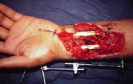
HIGH VELOCITY GUNSHOT WOUND IN THE FOREARM

HERIDA MANO DORSO

Preoperative picture: palmar view of the wound.

HERIDA PALMAR

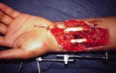
Preoperative picture: dorsal view of the wound.

COGAJO

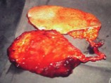
Picture of the free flap, in fact there are to flaps (latissimus dorsi and scapular) with one pedicle (subscapular vessels). The radial artery and comitant veins are used as recipient vessels; the latissimus dorsi muscle is used to cover the palm and the scapular skin the dorsum.

COLGAJO EN DORSO

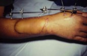
Four month postoperative picture, palmar view. (Grafted Latissimus Dorsi muscle flap).

COLGAJO EN PALMA

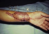
Four month postoperative picture, dorsal view. (Scapular skin flap).
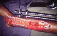
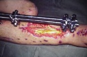
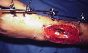

CLINICAL CASE Nº 2.

HERIDA Y AMPUTACION

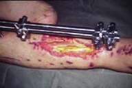
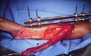
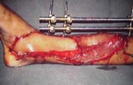
Picture 1. Left leg amputation and open fracture of right leg. It is very important to save the right leg, in order to rehabilitate the patient. 

RX FRACTURA

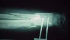
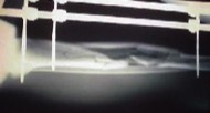
Picture 2. X Rays of the exposed tibia and fibula fracture.

ANTIPERSONNEL MINE IN THE LEGS

COLGAJO PIERNA
Picture 3. Late operative picture. After soft tissue reconstruction and open fracture coverage with Latissimus dorsi free flap and skin grafts.
POP COLGAJO

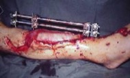
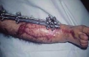
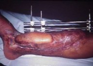
Picture 4. Postoperative picture 4 months after surgery.


CLINICAL CASE Nº 3.


FRAGMENTATION MISSILE IN THE LOWER EXTREMITY
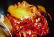
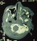
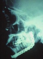
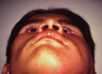
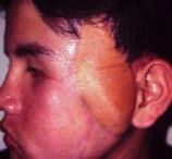
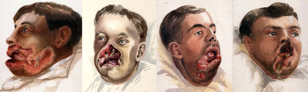
HERIDA GRANDE
Picture 1. Huge skin and muscle defect, with exposed tibia fracture and maleolar fracture.
COLGAJO LISTO
Picture 2. X Rays of the tibia fracture.
RX FRACTURA PIERNA
Picture 3. Latissimus dorsi free flap once it has been transferred, just before bone coverage.
COLGAJO PIERNA LISTO
Picture 4. Once the free flap has been completely sutured to cover both the tibia and maleolar fracture. The muscle is covered with skin grafts
COLGAJO POP
Picture 5. Five months postoperative picture.

CLINICAL CASE Nº 4.
HIGH VELOCITY GUNSHOT WOUND IN THE FACE

HERIDA CARA
Picture 1. Severe damage of soft tissues in the lateral aspect of the face. Destruction of skin, parotid gland, facial nerve, mandible, and zygoma.
TAC CARA
Picture 2. CT Scan shows the zygomatic arch and body destruction, mandible conminutive fracture with anterior displacement of the condile out of its place.
RX CARA
Picture 3. The zygoma body and zygomatic arch are reconstructed with a costal graft and AO microplates and screws. The mandible with an AO reconstruction plate from the condyle to the symphysis.
POP BASAL
Picture 4. The soft tissue defect was reconstructed with a parascupular free flap. The facial nerve was reconstructed with sural nerve grafts.
POP LATERAL
Picture 5. The zygoma is in acceptable position.

HERIDAS DE GUERRA EN LA CARA

dr bermudez en los archivos
GILLIES ARCHIVES OF FIRST WORLD WAR. WHEN PLASTIC SURGERY BEGUN.
One of the most amazing museums (private) I have ever visited were the Gillies Archives in the Queens Mary Hospital at Sidecup, UK.
I was able to review the medical records of the wounded treated by sir Harold Gillies during the first World War.
Here there are some of the images I took from their records showing the same wouds I have had to deal with in my country during the last decade.
Plastic surgery was born during first World War.
Complex wound in the extremities were treated by amputation, but there was a need to treat parts impossible to amputate : "face".
That was the reason why surgeons like Sir Harold Gillies needed to develop surgical techniques to reconstruct the wounded face.
fotos heridad 1 guerra mundial

Sus datos no son tomados sin su permiso.